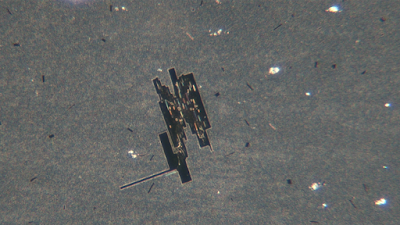
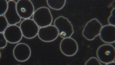
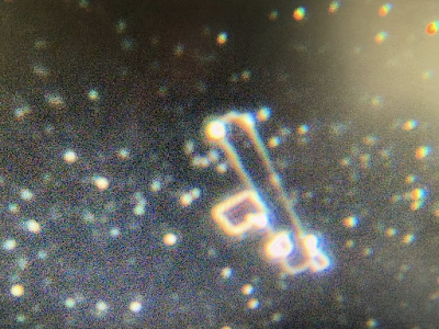
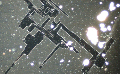
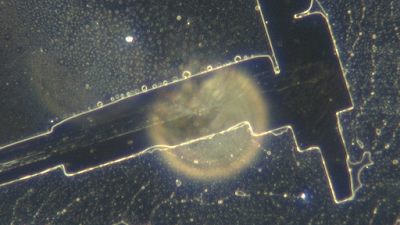
Masksaredangerous.com,29 January, 2022For those who just want the short story: Here’s the picture of one drop of New Zealand’s Pfizer COMIRNATY “vaccine” under a cover slip, after it was inadvertently heated lightly, and viewed the same day through dark field microscopy at low magnification, projected onto a TV monitor.
These images below are after a new computer with decent graphics was purchased along with software for the camera.
Increase the magnification on the image below and look closely.
The long story goes like this: I am a physician in good standing, having a background in mathematics and physics from university, then specialized beyond internal medicine after residency. I’ve had extensive phase contrast microscopy experience. People who were injured by the Pfizer jab started showing up in my practice. At this time in 2021, I didn’t have a microscope. Several doctors had come forward in other countries to report strange observations. Dr. Zandre Botha from South Africa showed the uniform strange round circles. La Quinta Columna showed what appeared to be microchips and other formations. Two other doctors talked about parasites and of all things, hydras! This is not my first rodeo. Having previously experienced how those of us that critically look at the vaccine situation are heavily infiltrated by persons wishing to divert and control the narrative, I thought, “What is real?”, “What is put forth in order to lead questioners to look like fools?” Because of my previous extensive microscopy experience, learning to use a dark field microscope didn’t take a huge amount of education. I took a 12 week course on live blood analysis using the most sophisticated dark field microscope and camera, that my money could buy. It magnifies up to 4000x. After having a close look at the blood of dozens of vaccine injured people, patients started asking me about certain round and square bright yellow formations in their capillary blood as seen on the screen.
Nothing in the textbooks identified such things, and various people doing similar work, suggested that they might be crystals or some sort of slide or cover slip anomaly. At first, I just told the patients that it wasn’t important—possibly uric acid or something like that. Then several “empty” vials of Pfizer COMIRNATY vaccine were obtained. I trust the source of the shots 100%. The vials were from recent leftover vaccine after the shots had been given. On a Friday in December, alone in my office, I took the vials out, thawed, and examined them. I drew up all the contents (just a few drops in each one) from each vial into a 3cc syringe with a 16G needle and put one drop of it on a slide with a cover slip, and another slide with a drop of vaccine mixed with a drop of human blood and put a cover slip on it. The images that follow are from the different slides and were taken immediately after the slides were made.
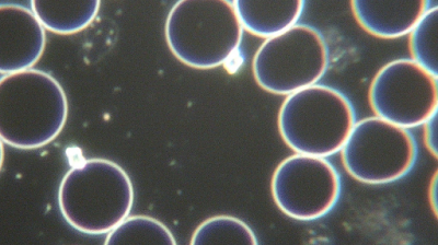
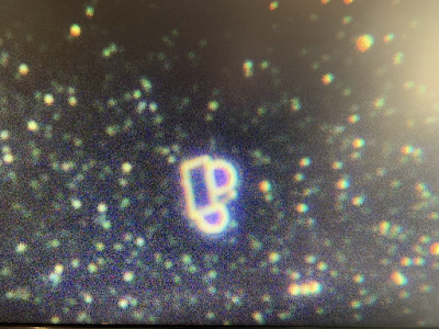
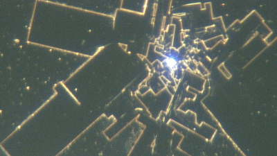
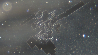
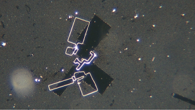
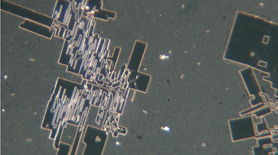
The amount of activity in the liquid and the strange shapes were baffling to say the least. Squares and circles seemed to connect up to each other consistently. Over an hour or two, the squares and circles seemed to enlarge somehow.
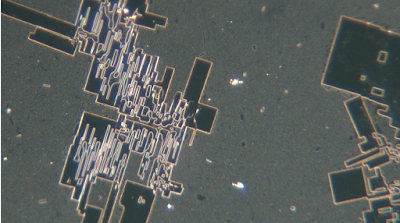
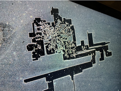
On the same hot day, I had to travel from my office to another office some distance away. The slides were packed up in plastic slide mailers and placed in a cardboard box in the car alongside the microscope. It had been a long week, and the ocean looked enticing, so I parked the car with the windows cracked about two inches, went for a 40-minute walk, then continued my drive. At my destination, the microscope was set up and I had another look at the slides. I don’t scare easily but what I saw next gave me a bit of a chill.
The straight lines and corners were lined with squares, rectangles, and circles Wanting better graphics, I purchased a gaming PC laptop. The software was then useable and I could take future images from the video program attached to the camera, for better definition. If this was to be believed by anyone, it had to be repeated and understood better. In the days that followed I contemplated how the formation happened. Was it a function of time? Of the vibration in the car? Heat? Cell towers along the way? It could have been any of those, so I took some more of the liquid and plated out some more slides, put them in multiple slide carriers, drove them all around the city near cell towers for two hours, and then came back. I did get some additional interesting images but nothing like the first one.
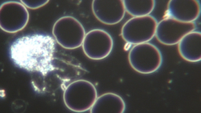
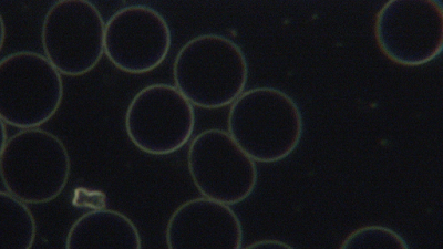
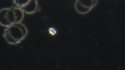
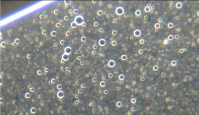
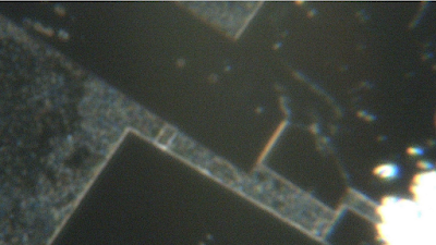
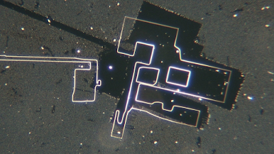
A few weeks later, I got hold of some more vaccine from the same source. This time there were lot numbers and four vials. I plated them out separately and also mixed some of the old liquid with the new. The new liquid had far more circles than the first lot, and had fewer squares.
The first attempts gave formations.
Interesting that on the second day when I came back and re-looked, the structures had shifted off their foundations. This may have been from handling the slides but it shows that the formations are “things” and not shadows or dehydration.
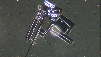
After some more attempts, interesting activity and organization occurred, but no dramatic new images were made.
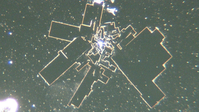
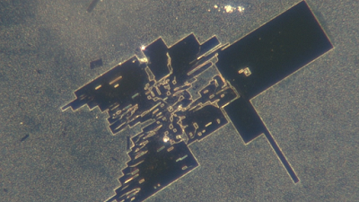
I plated out another batch on the 16th of January. Then I took them out in the car into the city on a hot day and left them in the car with the windows down. It was around 28°C outdoors, and the car got to around 40°C max. When I got home, this image was on the slide with a whole lot more. The experiment was successfully repeated.
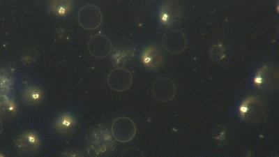
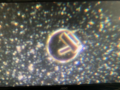
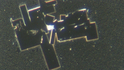
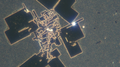
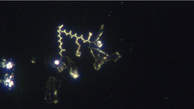
It is not for me to make assumptions on what these elements are, but I don’t think this is a normal finding in old style non-SARS-CoV-2 vaccines. The organisation that occurred does not appear to be simply organic, or debris, or artefact, and it is not simply dehydration of the slide. It is highly self-organised and appears to be part of the design of the liquid. For so many months I had pondered why this infection was so severe and important, that the vaccine has to be given to every human on the planet several times—even if one has recovered from Covid. This is unprecedented in vaccine history. Anyone with a dark field microscope can do this same thing. Why aren’t more people looking at the vaccine under a microscope? Why hasn’t the public been informed of the full ingredients of the only FDA approved vaccine for Covid? The following images are from the slide which had one drop of fresh human blood mixed with one drop of vaccine liquid. When the liquid met up to the blood, the white blood cells were annihilated, and the red blood cells heavily damaged. While concentrating on getting as good a focus as possible, I didn’t think too much of what I captured. But when I later looked closer, it was obviously an image of some sort of nano-tech chips linked by “cords”. The cords may be made in part from fibrin. But what is certain is that when mixed with human blood, different things than just “vaccine alone”, happen on the slide.
Have a look.
There were also these startling formations:
I have nothing else to say. The pictures speak for themselves.
This research should be continued by other laboratories.
Get some high quality German slides and cover slips. Place a drop of vaccine from a used vial or a fresh vial. Observe the activity at lower magnification and also at 1600 to 4000x using dark field microscopy. Get a dry incubator for the fresh slides and set it at different temperatures ranging from 38-42 for at least an hour. Observe again. If nothing interesting occurred, try putting it back in the incubator. Follow up the second day. Do the same with a drop of blood and a drop of vaccine. Combine different vaccine lots and use one drop of the combined vaccines on each slide.
Use PPE and vent the space so you are not breathing the liquid (a laminar flow hood would be ideal) and wear gloves
No comments:
Post a Comment
Note: only a member of this blog may post a comment.


No comments:
Post a Comment
Note: only a member of this blog may post a comment.